Groundbreaking Development: Human Cells Transformed into Minuscule Living Robots

Scientists have developed minuscule living robots using human cells, capable of autonomous movement in a laboratory dish This groundbreaking study suggests their potential in wound healing and tissue regeneration
Subscribe to CNN's Wonder Theory science newsletter for the latest news on remarkable discoveries, scientific breakthroughs, and more. In a recent study, researchers have successfully engineered miniature living robots using human cells, capable of mobility in a controlled environment. These tiny robots show promise in potential applications for wound healing and tissue regeneration in the future.
Researchers from Tufts University and Harvard University's Wyss Institute have named these innovative creations "anthrobots." This research project is an extension of the previous work done by the same team of scientists, who were the pioneers in creating the first living robots, known as xenobots, using stem cells extracted from embryos of the African clawed frog (Xenopus laevis).
"Some individuals believed that the characteristics of the xenobots were heavily dependent on the fact that they originated from embryonic and amphibian sources," stated Michael Levin, a co-author of the study and the Vannevar Bush Professor of Biology at Tufts School of Arts & Sciences.
"I believe this is not exclusive to embryos or frogs, but rather a common trait among all living organisms," he stated.
"We often underestimate the capabilities of our own body cells."
The anthrobots were considered incomplete organisms during their existence because they lacked a complete life cycle, according to Levin. "This serves as a reminder that the strict categorization of being either a robot, animal, or machine is not beneficial to us. We need to move past these limitations."
The research was published Thursday in the journal Advanced Science.
Gizem Gumuskaya is a doctoral student at Tufts University who helped create the anthrobots.
Gizem Gumuskaya, Tufts University
The scientists created them by using adult human cells from the trachea, or windpipe, obtained from anonymous donors of various ages and genders. This type of cell was chosen due to its accessibility and the belief that it had the potential for motion, which was important for their research on Covid-19 and lung disease, as explained by study coauthor Gizem Gumuskaya, a doctoral student at Tufts.
The tracheal cells are equipped with cilia, which are hairlike projections that wave back and forth. These cilia are responsible for assisting the tracheal cells in expelling small particles that may enter the air passages of the lungs. Previous research has also demonstrated the cells' ability to form organoids, which are clumps of cells commonly utilized for research.
Cao et al./Courtesy Cell
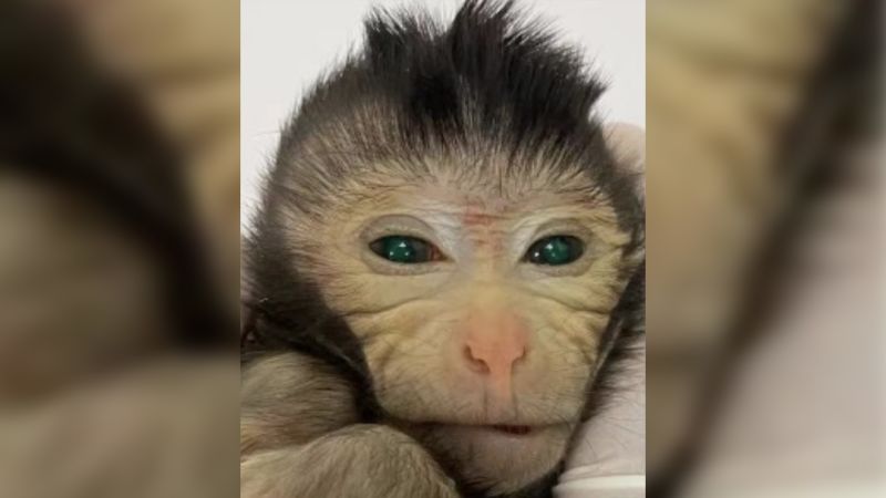
Scientists create chimeric monkey with two sets of DNA
Gumuskaya conducted experiments on the chemical composition of tracheal cell growth conditions and successfully discovered a method to promote the outward orientation of cilia on the organoids. After identifying the optimal matrix, the organoids exhibited mobility within a few days, with the cilia functioning similarly to oars.
"On day one, two, four, and five, there were no changes, but around day seven, as is typical in biology, a rapid transformation occurred," she explained. "It was akin to a flower blooming. By day seven, the cilia had reversed and were positioned on the exterior."
Each anthrobot in our method originates from a single cell, making them unique. While other scientists have created biological robots, they were manually constructed using molds and seeding cells, according to Levin.
Each anthrobot grows from a single cell.
Gizem Gumuskaya, Tufts University
Different shapes and sizes
The team's created anthrobots were not all the same. Some were spherical and completely covered in cilia, while others had a football-like shape with irregular cilia coverage. They also exhibited various movement patterns, including straight lines, tight circles, and some simply sat around and wiggled, as reported in a study news release. These anthrobots were able to survive for up to 60 days in laboratory conditions.
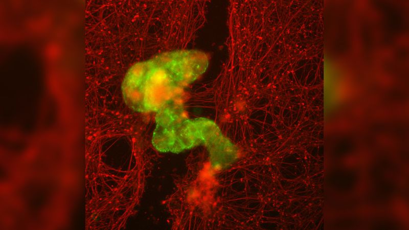
The experiments described in the recent study are still in the early stages, with the objective being to determine the potential medical uses of anthrobots, stated Levin and Gumuskaya. In order to investigate the possible applications, scientists studied the ability of anthrobots to travel over human neurons that had been artificially damaged in a laboratory setting. The study revealed that the anthrobots were able to stimulate growth in the damaged areas of the neurons, although the exact mechanism of healing is not yet understood by the researchers.
Falk Tauber, a group leader at the Freiburg Center for Interactive Materials and Bioinspired Technologies at the University of Freiburg in Germany, emphasized that the study has laid the groundwork for future endeavors to utilize the bio-bots for various purposes and to create them in diverse configurations.
An anthrobot, in green, grows across a scratch through neuronal tissue, in red.
Gizem Gumuskaya, Tufts University
Tauber, not part of the study, observed that the anthrobots displayed unexpected behavior, especially as they moved and eventually closed scratches in human neurons. He mentioned that the capability to create these structures from a patient's own cells has various potential applications in the lab and potentially in humans.
Levin expressed that he had no ethical or safety concerns regarding the anthrobots. According to him, they are not created from human embryos, genetically modified, or subjected to restricted research.
He added that the anthrobots have a limited environment and cannot survive outside the lab. Additionally, they have a natural lifespan and seamlessly biodegrade after a few weeks.


